What is Retinal Imaging?
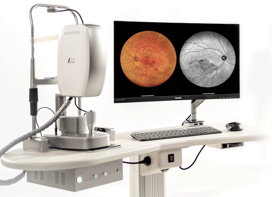
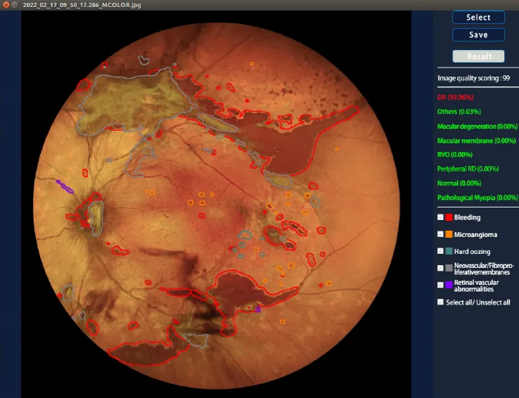
Retinal imaging is a diagnostic test that creates high-quality digital images of the inner, back surface of your eye. It allows the diagnosis of many eye conditions like diabetes-related retinopathy, glaucoma and macular degeneration. Your provider will tell you how often you need retinal imaging.

What is retinal imaging?
In retinal imaging, low-power lasers or a camera flash emit light into the eye through the pupil. The reflected light exits through the same path and is captured by the device, generating an image of the retina. While similar imaging techniques are employed during routine eye health check-ups at mainstream optometry practices, our analysis delves deeper into these images, seeking additional insights into the overall health of the human body and brain.
What it used for?
The back of the eye is known as the retina, offering a unique and easily observable view of blood vessels and nerves, unlike many other parts of the human body. While these anatomical structures are also present in the brain, accessing them is considerably more challenging. Our research focuses on leveraging information obtained from retinal images to gain insights into brain activity.
Subtle changes in the retina may mirror similar processes occurring in the brain, and these early indicators could precede declines in brain health by several years or even decades. Additionally, examining blood vessels in the eye proves valuable in identifying and comprehending diseases affecting the human circulatory system, such as high blood pressure, diabetes, and heart disease.
For instance, through further investigation, we may soon be able to detect individuals with undiagnosed high blood pressure by analyzing images of their retina. This advancement could empower healthcare professionals to prescribe suitable medications, significantly reducing the risk of future heart attacks or strokes.

Retinal imaging can be used to see:
- Blood vessels that lie close to the surface as well as those that are located deeper in the retina
- The optic nerve head where blood vessels and nerves enter and leave the eye
- The different cellular layers which contain nerves and axons
- The macular, which is the part of our eye responsible for central vision
How is retinal imaging performed?
Eye care specialists employ three primary methods to capture digital images of the fundus, the inner back surface of the eye. These methods include:
1. Color fundus photos: Fundus, referring to the back of the eye, is captured through the use of fundus cameras, a technique employed for decades. During the process, bright flashes of light are used to take pictures, and technological advancements now enable precise digital retinal images in high resolution. Some cameras can provide wide-field views of the fundus, allowing the eye care team to examine a larger area of the retina. This imaging method excels in revealing blood vessels and indicating signs of diabetes-related retinopathy.
2. Optical coherence tomography (OCT): A common imaging test, OCT offers cross-sectional views of the layers in the macula region and optic nerve. This method provides detailed information about each retinal layer's thickness, aiding in the diagnosis of conditions such as diabetes-related macular edema and macular degeneration.
In all instances, you sit comfortably in a chair and bring your face close to the camera device. Your healthcare provider guides you on placing your forehead and chin in the designated positions. Importantly, there is no contact with your eye during the retinal imaging process.
Providers may employ one or a combination of the mentioned methods simultaneously. Additionally, they might incorporate fluorescein angiography in conjunction with one of the methods. This minimally invasive technique involves injecting a dye into a vein in your arm. The dye circulates through your blood vessels, including those in your eyes, revealing any blockages or other issues in these vessels. Notably, this procedure does not involve any direct contact with your eyes.
More details please visit www.microcleartech.net
None

Comments